Customers

Our certified sonographers deliver accurate, on-site diagnostic imaging across a comprehensive range of ultrasound specialties. Select a service below to learn more.

Mobile to Your Location

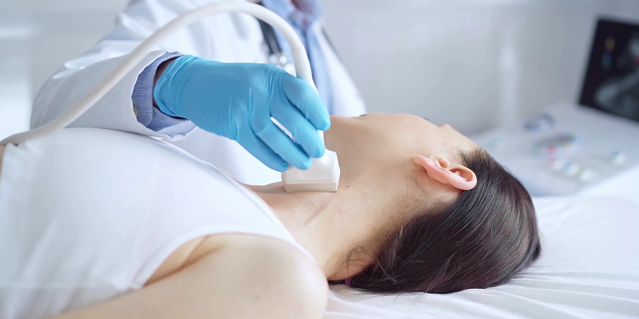
We bring advanced ultrasound imaging directly to hospitals, skilled nursing facilities, physician offices, assisted living communities, and patient homes across the Charlotte metro area.

Board-Certified Radiologist Reports

Every study is interpreted by a board-certified radiologist with rapid turnaround — typically within hours — so referring providers can act on results without delay.

Same-Day & STAT Service

Our ARDMS-certified sonographers are available seven days a week, including same-day and STAT requests, to support urgent clinical needs whenever they arise.

Diagnostic Ultrasound Services


Abdominal Ultrasound

Abdominal ultrasound anatomical illustration

An abdominal ultrasound delivers detailed, real-time visualization of the body’s core organs — including the liver, gallbladder, pancreas, spleen, and kidneys. This painless, 30-minute examination helps physicians identify a wide range of conditions without the need for invasive procedures. Patients should refrain from eating or drinking for 6 to 8 hours prior to the exam.

Common Indications

  • Persistent Abdominal Pain
  • Nausea or Vomiting
  • Suspected Gallstones
  • Jaundice or Elevated Bilirubin
  • Abnormal Liver Enzymes
  • Fluid Accumulation in the Abdomen
  • Fatty Liver Disease
  • Unexplained Gas or Bloating
  • Urinary Tract Obstruction

Diagnostic Findings

  • Gallbladder Stones
  • Kidney Stones or Masses
  • Liver Disease including Cirrhosis
  • Pancreatic Abnormalities
  • Splenic Enlargement
  • Abdominal Aortic Aneurysm
  • Hydronephrosis
  • Organ Cysts or Tumors

Arterial Ultrasound

Arterial ultrasound lower extremity illustration

Peripheral arterial disease affects millions of adults, particularly those over 50 with risk factors like diabetes or tobacco use. Our arterial ultrasound evaluates blood flow through the upper and lower extremities to detect plaque buildup, narrowing, or blockages before they progress to serious complications. The exam is completely noninvasive and typically completed within 30 minutes.

Common Indications

  • Leg or Arm Pain During Activity
  • Pain at Rest in the Extremities
  • History of Diabetes
  • Tobacco Use
  • Non-Healing Wounds or Ulcers
  • Pre- or Post-Surgical Evaluation
  • Known Abdominal Aneurysm
  • Intermittent Claudication

Diagnostic Findings

  • Peripheral Arterial Disease
  • Arterial Stenosis or Blockage
  • Popliteal Aneurysm
  • Baker’s Cyst
  • Vascular Insufficiency

Carotid Ultrasound

Carotid ultrasound neck anatomical illustration

Carotid ultrasound is the primary screening tool for evaluating the arteries that supply blood to the brain. Given that cardiovascular disease and stroke remain among the top causes of death in the U.S., early detection of arterial narrowing is critical. This quick, in-office exam takes roughly 30 minutes and is recommended for patients with key risk factors such as high blood pressure, elevated cholesterol, or a family history of vascular disease.

Common Indications

  • Episodes of Slurred Speech
  • Temporary Vision Loss
  • Unexplained Dizziness or Vertigo
  • Loss of Coordination
  • Fainting or Near-Fainting
  • Detectable Carotid Bruit
  • Pre-Operative Screening
  • Speech Difficulties

Diagnostic Findings

  • Carotid Artery Narrowing
  • Arterial Wall Thickening
  • Risk of Transient Ischemic Attack
  • Stroke Risk Assessment
  • Compromised Cerebral Blood Flow
  • Subclavian Artery Stenosis

Echocardiogram Ultrasound

Echocardiogram heart anatomical illustration

An echocardiogram uses ultrasound technology to produce a moving picture of the heart, allowing physicians to evaluate how effectively it beats, pumps blood, and manages flow through its chambers and valves. This examination is essential for diagnosing structural and functional cardiac conditions and plays a vital role in ongoing cardiovascular care.

Common Indications

  • Chest Pain or Discomfort
  • Persistent Shortness of Breath
  • Severe or Resistant Hypertension
  • Heart Murmur Detection
  • Irregular Heart Rhythm
  • Fainting or Near-Syncope Episodes
  • History of Congestive Heart Failure
  • Suspected Atrial Fibrillation

Diagnostic Findings

  • Valve Disorders and Regurgitation
  • Aortic Valve Stenosis
  • Heart Muscle Weakness
  • Congenital Cardiac Defects
  • Fluid Around the Heart
  • Pulmonary Hypertension
  • Prior Heart Attack Damage
  • Overall Cardiac Function

Pelvic Ultrasound

Pelvic ultrasound anatomical illustration

Pelvic ultrasound offers a safe and reliable method for evaluating the reproductive and urinary organs, including the uterus, ovaries, fallopian tubes, cervix, and bladder. Using Doppler technology to assess blood flow patterns, this 30-minute exam provides critical diagnostic information. A full bladder is typically recommended for optimal imaging.

Common Indications

  • Pelvic Pain or Pressure
  • Suspected Fibroids
  • Abnormal or Irregular Bleeding
  • Missed or Absent Menstrual Periods
  • Pelvic Inflammatory Concerns
  • Endometrial Thickening
  • Urinary Tract Obstruction
  • Post-Menopausal Bleeding
  • Ovarian Mass Evaluation

Diagnostic Findings

  • Uterine Fibroids
  • Ovarian Cysts or Masses
  • Endometriosis
  • Ectopic Pregnancy
  • Pelvic Inflammatory Disease
  • Ovarian Torsion
  • Dermoid Cysts
  • Cervical or Bladder Abnormalities

Renal Ultrasound

Renal ultrasound kidney anatomical illustration

A renal ultrasound provides a thorough, noninvasive assessment of the kidneys, ureters, and bladder. Using real-time imaging enhanced by color Doppler, this exam identifies structural abnormalities, fluid blockages, stones, and masses within the urinary system. No special preparation is needed, and results are typically available in under 30 minutes.

Common Indications

  • Flank or Lower Back Pain
  • Blood in the Urine
  • Suspected Kidney Stones
  • Elevated Protein in Urine
  • Swelling of the Kidneys
  • Recurrent Bladder Infections
  • Kidney Infection Symptoms

Diagnostic Findings

  • Kidney Stone Location and Size
  • Hydronephrosis
  • Renal Cysts or Solid Masses
  • Ureteral Dilation
  • Incomplete Bladder Emptying
  • Renal Cell Carcinoma
  • Acute Kidney Injury

Scrotal Ultrasound

Scrotal ultrasound anatomical illustration

Scrotal ultrasound is widely regarded as the definitive imaging method for evaluating testicular health. This 30-minute examination provides high-resolution, real-time images of the testicles, epididymis, spermatic cord, and surrounding structures while simultaneously monitoring arterial and venous blood flow to detect a broad range of conditions.

Common Indications

  • Testicular Pain or Swelling
  • Palpable Scrotal Mass
  • Suspected Testicular Torsion
  • Varicocele Symptoms
  • Epididymal Inflammation
  • Spermatocele Evaluation
  • Hydrocele Assessment
  • Unexplained Scrotal Discomfort

Diagnostic Findings

  • Testicular Torsion
  • Testicular Cancer
  • Epididymitis
  • Varicocele
  • Spermatocele
  • Hydrocele
  • Orchitis

Thyroid Ultrasound

Thyroid ultrasound neck anatomical illustration

The thyroid gland, located at the base of the neck, produces hormones that govern metabolism, energy levels, heart rate, and body temperature. A thyroid ultrasound provides a fast, painless assessment of the gland’s size, shape, and internal structure, making it an essential tool for evaluating nodules, inflammation, and other thyroid disorders. The exam is typically completed in under 30 minutes.

Common Indications

  • Overactive or Underactive Thyroid
  • Palpable Neck Mass or Nodule
  • Abnormal Thyroid Lab Results
  • Difficulty Swallowing
  • Thyroid Inflammation
  • Follow-Up on Known Nodules
  • Enlarged Thyroid Gland

Diagnostic Findings

  • Thyroid Nodules or Cancer
  • Goiter
  • Tracheal Compression
  • Parathyroid Masses
  • Lymph Node Enlargement

Venous Ultrasound

Venous ultrasound lower extremity illustration

Venous ultrasound examines the deep and superficial veins of the upper or lower extremities to evaluate circulation and detect obstructions. This study is the frontline diagnostic tool for identifying blood clots and assessing chronic venous insufficiency, which can lead to varicose veins and other complications. The exam requires no preparation and is completed in under 30 minutes.

Common Indications

  • Leg Pain or Heaviness
  • Swelling in the Extremities
  • Unexplained Edema
  • Sudden Shortness of Breath
  • Suspected Deep Vein Thrombosis
  • Varicose Vein Evaluation

Diagnostic Findings

  • Deep Vein Thrombosis
  • Superficial Blood Clots
  • Baker’s Cyst
  • Chronic Venous Insufficiency
  • Venous Reflux

Other Specialty Ultrasound Studies

Specialty ultrasound illustration

Beyond our standard catalog of studies, our sonographers are credentialed to perform additional specialty exams on request. If you need a study that isn’t listed on the tabs above, contact us — we will work with you to confirm protocol, scheduling, and reporting for the exam you need. This flexibility lets your facility offer a broader imaging program without expanding equipment or staff.

Common Indications

  • Resistant Hypertension
  • Known Atherosclerotic Disease
  • Age Over 50 with Risk Factors
  • Diabetes with Renal Concerns
  • Audible Abdominal Bruit
  • Decreased Kidney Size
  • Chronic Kidney Disease
  • Sudden Decline in Kidney Function

Diagnostic Findings

  • Renal Artery Stenosis
  • Progressive Kidney Failure
  • Renal Vein Blockage
  • Arterial Embolism
  • Kidney Masses or Cysts
  • Renal Carcinoma
  • Kidney Size Abnormalities

How Our Ultrasound Service Works

From request to radiologist report, we make mobile ultrasound simple for providers and patients.

1

Request a Study

Call, fax, or submit an order through our secure provider portal. Our team confirms details the same day.

2

Sonographer Dispatched

An ARDMS-certified sonographer is scheduled and dispatched to your facility or the patient’s location.

3

On-Site Ultrasound Exam

The study is performed bedside using hospital-grade portable ultrasound equipment — no patient transport required.

4

Radiologist Report Delivered

Images are transmitted to a board-certified radiologist, and the final report is delivered to the referring provider — often within hours.

Trusted by Providers Across the Charlotte Region

What referring physicians and facility partners say about working with Viason.

“Viason’s mobile ultrasound service has been a game-changer for our skilled nursing facility. Their sonographers are professional, reports come back quickly, and our residents never have to leave the building for imaging.”

Director of Nursing
Charlotte-area Skilled Nursing Facility

“Same-day turnaround on vascular studies has meaningfully improved our patient care. Viason is reliable, responsive, and the imaging quality is excellent.”

Primary Care Physician
Charlotte, NC

“We’ve partnered with Viason for years for echocardiograms and abdominal studies. Their team is consistently courteous with patients and their radiologist reports are thorough and timely.”

Office Manager
Independent Physician Practice

Our Ultrasound Service Area

Viason provides mobile diagnostic ultrasound throughout the greater Charlotte metropolitan area.

For more than 27 years, medical practices, hospitals, skilled nursing facilities, assisted living communities, and home-bound patients across the Charlotte region have relied on Viason for accurate, on-site diagnostic ultrasound imaging.

Counties served include:

  • Mecklenburg County, NC
  • Union County, NC
  • Cabarrus County, NC
  • Gaston County, NC
  • Iredell County, NC
  • Lincoln County, NC
  • York County, SC
  • Lancaster County, SC

Don’t see your location? Contact us — we may still be able to serve you.

Frequently Asked Questions

Common questions about our mobile ultrasound service.

How quickly can a sonographer be on-site?
For routine studies, we typically schedule within 24 hours. Same-day and STAT ultrasound requests are accommodated seven days a week based on clinical urgency.
What types of facilities do you serve?
We provide mobile ultrasound at hospitals, skilled nursing facilities, assisted living communities, physician offices, outpatient clinics, hospice settings, and private residences for home-bound patients.
Are your sonographers credentialed?
Yes. All of our sonographers are ARDMS-certified (American Registry for Diagnostic Medical Sonography) and experienced in performing the full range of diagnostic ultrasound studies we offer.
Who reads the ultrasound images?
Every study is interpreted by a board-certified radiologist. Final reports are delivered to the referring provider — typically within hours of the exam being completed.
How is billing handled?
Viason bills Medicare, Medicaid, and most commercial insurance plans directly. Our billing team can answer questions about coverage and out-of-pocket costs prior to scheduling.
Do you offer after-hours or weekend ultrasound service?
Yes. We operate seven days a week and can accommodate after-hours and weekend requests for urgent diagnostic needs.
How do I order an ultrasound study?
Referring providers can place orders by phone, fax, or through our secure provider portal. Contact us to get set up as a referring provider.

Schedule a Mobile Ultrasound Today

Our team is standing by to coordinate same-day, next-day, and STAT ultrasound studies throughout the Charlotte region.

Contact Viason