Patient Resources
Everything you need to know about your upcoming ultrasound — what to expect, how to prepare, and what happens after your exam. We bring hospital-grade diagnostic imaging right to your physician's office, so you can focus on what matters most.
30 Min Average Exam
No Radiation
In Your Doctors Office
Same-Day Prelim Results
What Is an Ultrasound?
An ultrasound is a safe, painless, non-invasive diagnostic exam that uses high-frequency sound waves — not radiation — to produce real-time images of soft tissues, blood vessels, and organs inside the body. Sound waves are reflected by tissue and converted into images by a sophisticated computer system, allowing your physician to evaluate your health without any incisions or injections.
Because ultrasound uses sound rather than radiation, it is considered one of the safest imaging modalities available — appropriate for patients of all ages, including pregnant women.
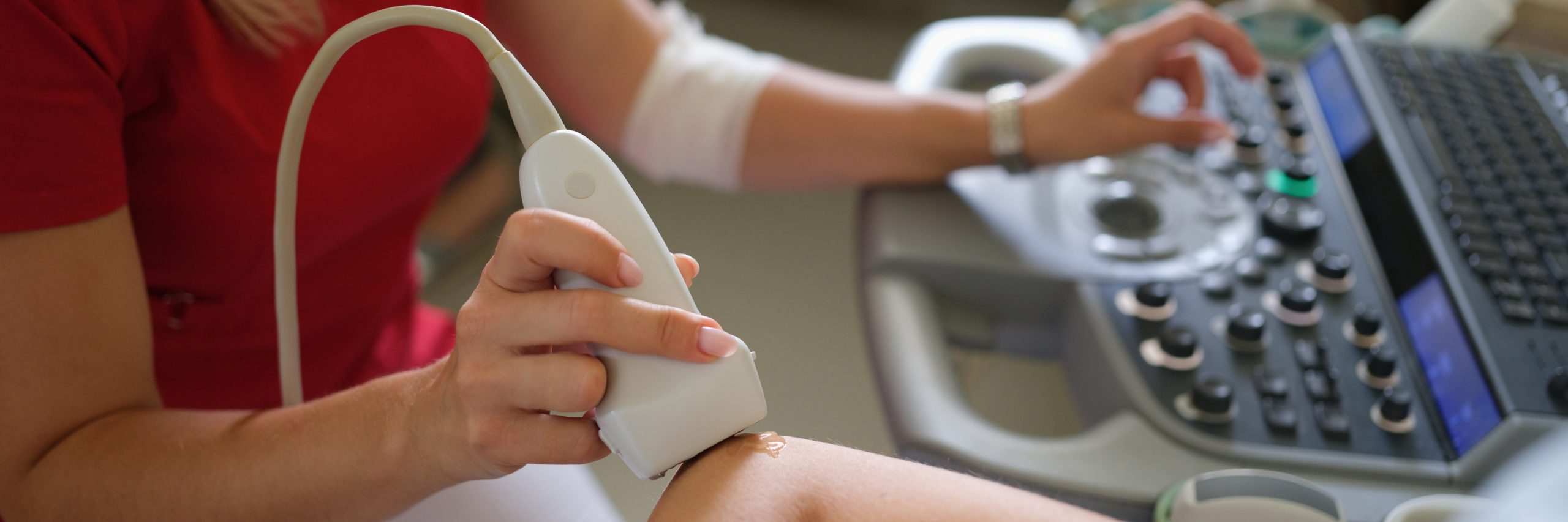
What to Expect During Your Exam
Your ultrasound is straightforward, comfortable, and typically completed in 30 minutes or less.
1
Get Comfortable
You’ll be positioned on an exam table — typically on your back or side, depending on the study.
2
Gel Applied
A warm, water-soluble gel is applied to the area being scanned to help the sound waves travel.
3
Imaging Begins
The sonographer moves a transducer (handheld probe) across your skin, capturing real-time images.
4
Stay Still or Hold Breath
You may be asked to hold your breath briefly or remain very still to capture clear images.
5
All Done
The gel is wiped off, you can resume normal activities immediately. No recovery needed.
Our Diagnostic Ultrasound Services
Learn more about your specific exam — common indications, diagnostic findings, and what each study evaluates.
Abdominal Ultrasound
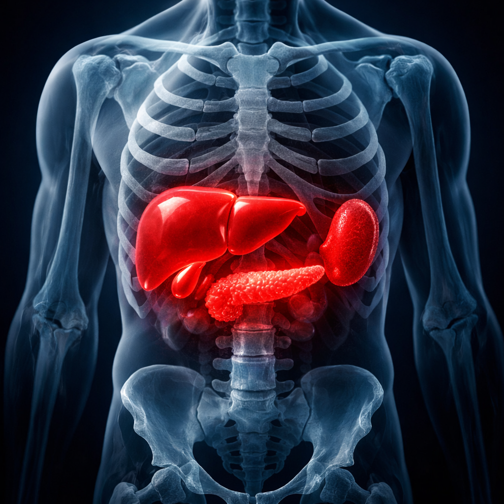
An abdominal ultrasound delivers detailed, real-time visualization of the body’s core organs — including the liver, gallbladder, pancreas, spleen, and kidneys. This painless, 30-minute examination helps physicians identify a wide range of conditions without the need for invasive procedures. Patients should refrain from eating or drinking for 6 to 8 hours prior to the exam.
Common Indications
- Persistent Abdominal Pain
- Nausea or Vomiting
- Suspected Gallstones
- Jaundice or Elevated Bilirubin
- Abnormal Liver Enzymes
- Fluid Accumulation in the Abdomen
- Fatty Liver Disease
- Unexplained Gas or Bloating
- Urinary Tract Obstruction
Diagnostic Findings
- Gallbladder Stones
- Kidney Stones or Masses
- Liver Disease including Cirrhosis
- Pancreatic Abnormalities
- Splenic Enlargement
- Abdominal Aortic Aneurysm
- Hydronephrosis
- Organ Cysts or Tumors
Arterial Ultrasound
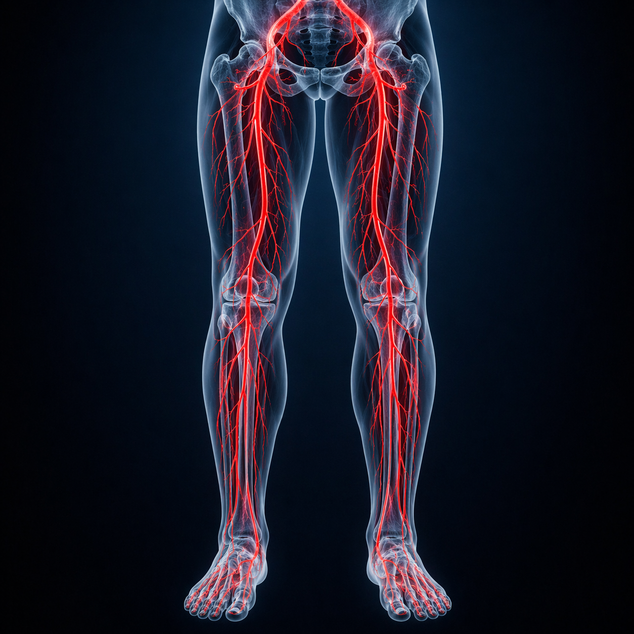
Peripheral arterial disease affects millions of adults, particularly those over 50 with risk factors like diabetes or tobacco use. Our arterial ultrasound evaluates blood flow through the upper and lower extremities to detect plaque buildup, narrowing, or blockages before they progress to serious complications. The exam is completely noninvasive and typically completed within 30 minutes.
Common Indications
- Leg or Arm Pain During Activity
- Pain at Rest in the Extremities
- History of Diabetes
- Tobacco Use
- Non-Healing Wounds or Ulcers
- Pre- or Post-Surgical Evaluation
- Known Abdominal Aneurysm
- Intermittent Claudication
Diagnostic Findings
- Peripheral Arterial Disease
- Arterial Stenosis or Blockage
- Popliteal Aneurysm
- Baker’s Cyst
- Vascular Insufficiency
Carotid Ultrasound
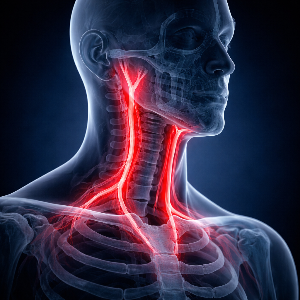
Carotid ultrasound is the primary screening tool for evaluating the arteries that supply blood to the brain. Given that cardiovascular disease and stroke remain among the top causes of death in the U.S., early detection of arterial narrowing is critical. This quick, in-office exam takes roughly 30 minutes and is recommended for patients with key risk factors such as high blood pressure, elevated cholesterol, or a family history of vascular disease.
Common Indications
- Episodes of Slurred Speech
- Temporary Vision Loss
- Unexplained Dizziness or Vertigo
- Loss of Coordination
- Fainting or Near-Fainting
- Detectable Carotid Bruit
- Pre-Operative Screening
- Speech Difficulties
Diagnostic Findings
- Carotid Artery Narrowing
- Arterial Wall Thickening
- Risk of Transient Ischemic Attack
- Stroke Risk Assessment
- Compromised Cerebral Blood Flow
- Subclavian Artery Stenosis
Echocardiogram Ultrasound
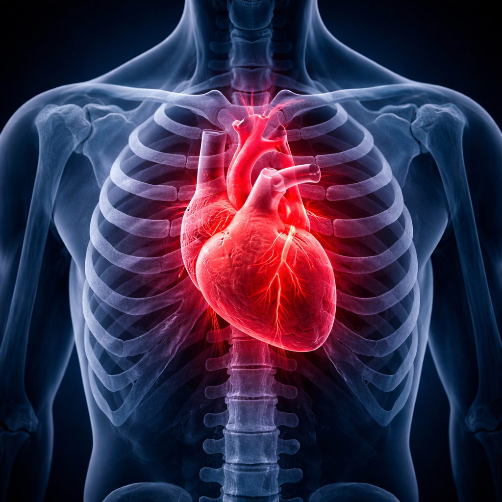
An echocardiogram uses ultrasound technology to produce a moving picture of the heart, allowing physicians to evaluate how effectively it beats, pumps blood, and manages flow through its chambers and valves. This examination is essential for diagnosing structural and functional cardiac conditions and plays a vital role in ongoing cardiovascular care.
Common Indications
- Chest Pain or Discomfort
- Persistent Shortness of Breath
- Severe or Resistant Hypertension
- Heart Murmur Detection
- Irregular Heart Rhythm
- Fainting or Near-Syncope Episodes
- History of Congestive Heart Failure
- Suspected Atrial Fibrillation
Diagnostic Findings
- Valve Disorders and Regurgitation
- Aortic Valve Stenosis
- Heart Muscle Weakness
- Congenital Cardiac Defects
- Fluid Around the Heart
- Pulmonary Hypertension
- Prior Heart Attack Damage
- Overall Cardiac Function
Pelvic Ultrasound
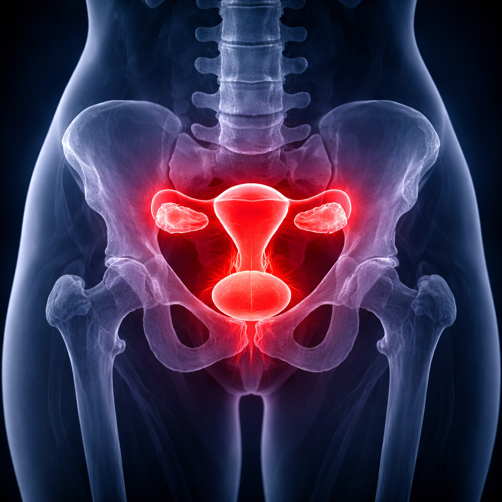
Pelvic ultrasound offers a safe and reliable method for evaluating the reproductive and urinary organs, including the uterus, ovaries, fallopian tubes, cervix, and bladder. Using Doppler technology to assess blood flow patterns, this 30-minute exam provides critical diagnostic information. A full bladder is typically recommended for optimal imaging.
Common Indications
- Pelvic Pain or Pressure
- Suspected Fibroids
- Abnormal or Irregular Bleeding
- Missed or Absent Menstrual Periods
- Pelvic Inflammatory Concerns
- Endometrial Thickening
- Urinary Tract Obstruction
- Post-Menopausal Bleeding
- Ovarian Mass Evaluation
Diagnostic Findings
- Uterine Fibroids
- Ovarian Cysts or Masses
- Endometriosis
- Ectopic Pregnancy
- Pelvic Inflammatory Disease
- Ovarian Torsion
- Dermoid Cysts
- Cervical or Bladder Abnormalities
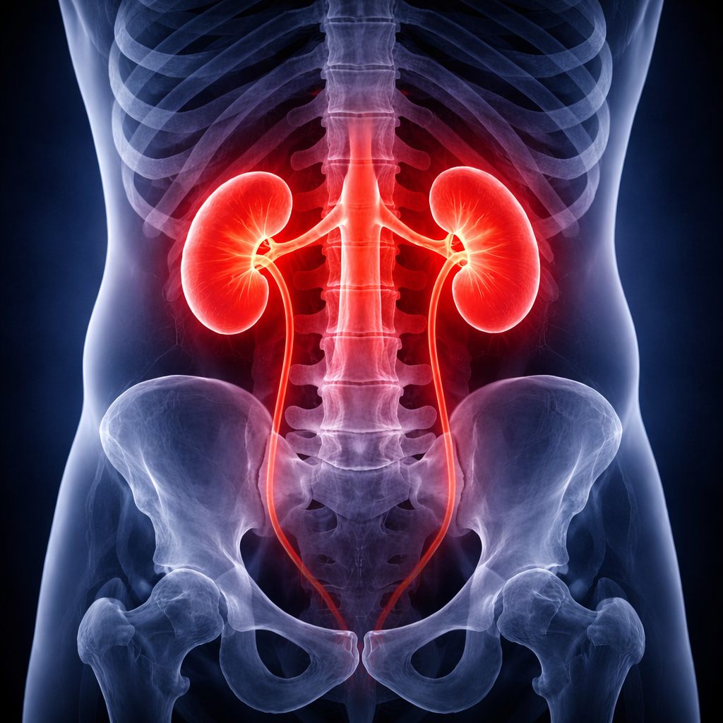
Renal Ultrasound
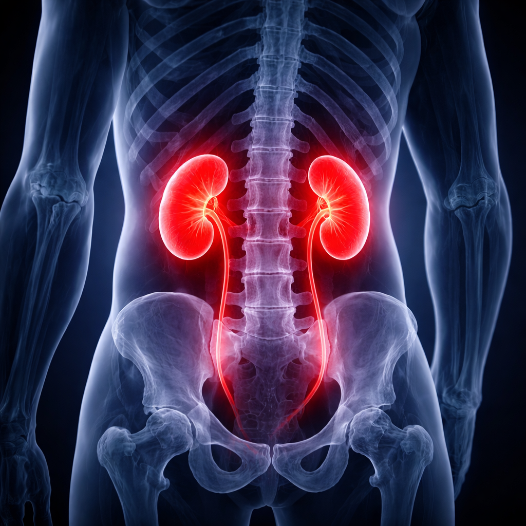
A renal ultrasound provides a thorough, noninvasive assessment of the kidneys, ureters, and bladder. Using real-time imaging enhanced by color Doppler, this exam identifies structural abnormalities, fluid blockages, stones, and masses within the urinary system. No special preparation is needed, and results are typically available in under 30 minutes.
Common Indications
- Flank or Lower Back Pain
- Blood in the Urine
- Suspected Kidney Stones
- Elevated Protein in Urine
- Swelling of the Kidneys
- Recurrent Bladder Infections
- Kidney Infection Symptoms
Diagnostic Findings
- Kidney Stone Location and Size
- Hydronephrosis
- Renal Cysts or Solid Masses
- Ureteral Dilation
- Incomplete Bladder Emptying
- Renal Cell Carcinoma
- Acute Kidney Injury
Scrotal Ultrasound
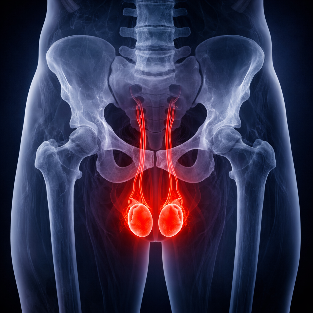
Scrotal ultrasound is widely regarded as the definitive imaging method for evaluating testicular health. This 30-minute examination provides high-resolution, real-time images of the testicles, epididymis, spermatic cord, and surrounding structures while simultaneously monitoring arterial and venous blood flow to detect a broad range of conditions.
Common Indications
- Testicular Pain or Swelling
- Palpable Scrotal Mass
- Suspected Testicular Torsion
- Varicocele Symptoms
- Epididymal Inflammation
- Spermatocele Evaluation
- Hydrocele Assessment
- Unexplained Scrotal Discomfort
Diagnostic Findings
- Testicular Torsion
- Testicular Cancer
- Epididymitis
- Varicocele
- Spermatocele
- Hydrocele
- Orchitis
Thyroid Ultrasound
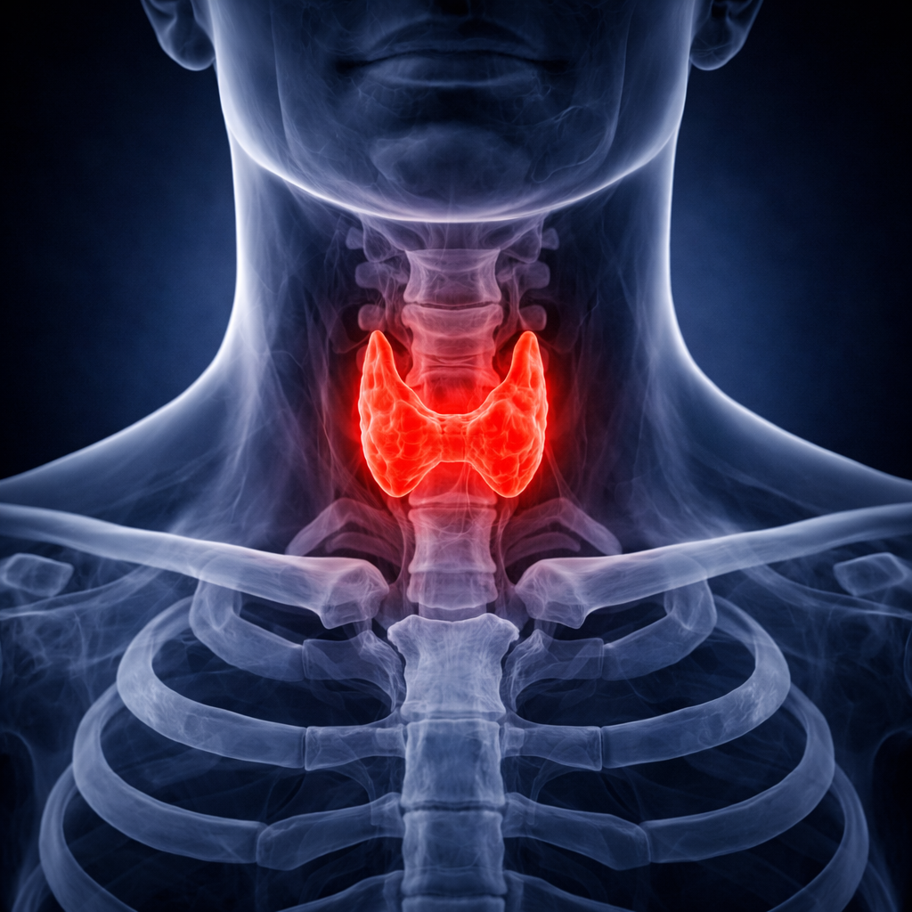
The thyroid gland, located at the base of the neck, produces hormones that govern metabolism, energy levels, heart rate, and body temperature. A thyroid ultrasound provides a fast, painless assessment of the gland’s size, shape, and internal structure, making it an essential tool for evaluating nodules, inflammation, and other thyroid disorders. The exam is typically completed in under 30 minutes.
Common Indications
- Overactive or Underactive Thyroid
- Palpable Neck Mass or Nodule
- Abnormal Thyroid Lab Results
- Difficulty Swallowing
- Thyroid Inflammation
- Follow-Up on Known Nodules
- Enlarged Thyroid Gland
Diagnostic Findings
- Thyroid Nodules or Cancer
- Goiter
- Tracheal Compression
- Parathyroid Masses
- Lymph Node Enlargement
Venous Ultrasound
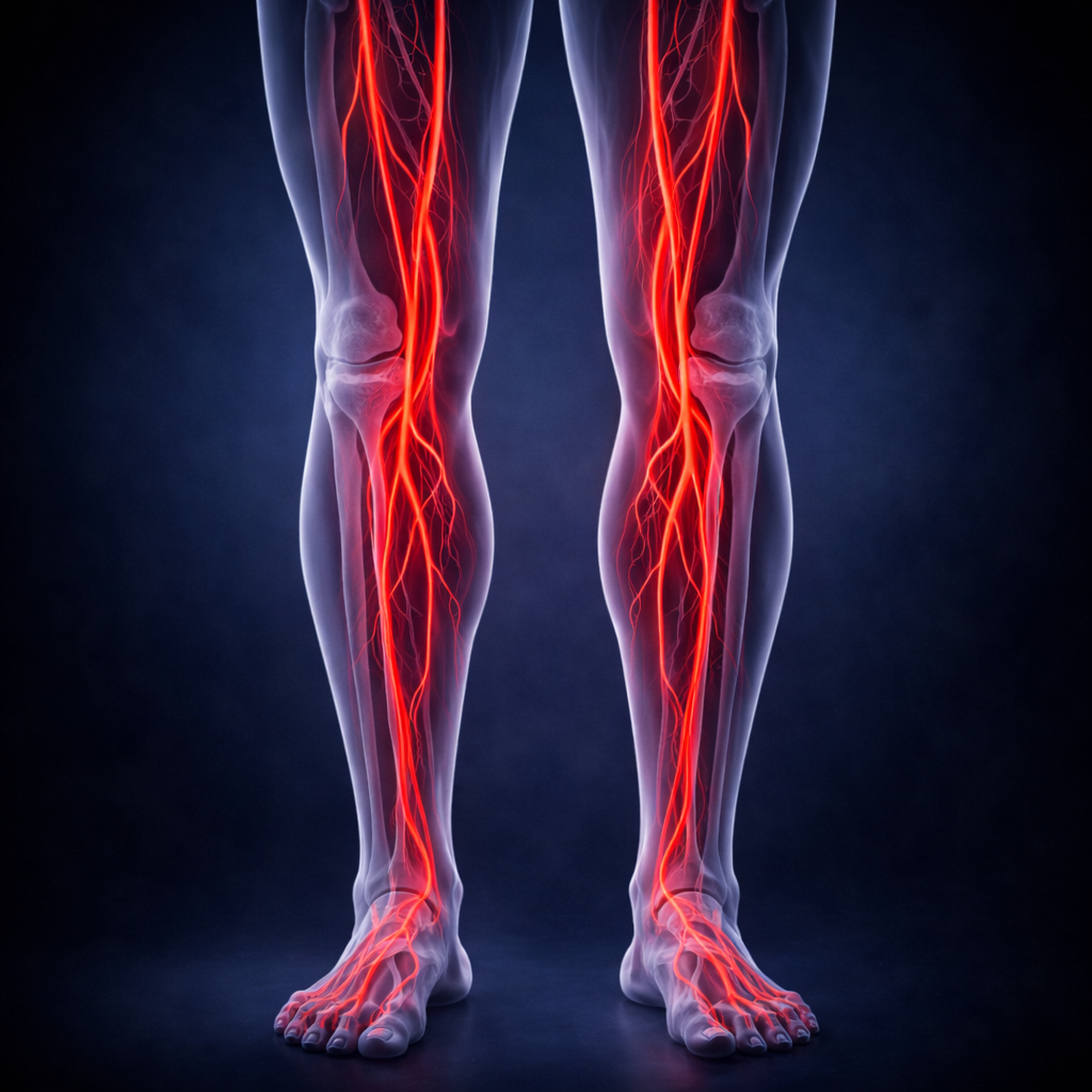
Venous ultrasound examines the deep and superficial veins of the upper or lower extremities to evaluate circulation and detect obstructions. This study is the frontline diagnostic tool for identifying blood clots and assessing chronic venous insufficiency, which can lead to varicose veins and other complications. The exam requires no preparation and is completed in under 30 minutes.
Common Indications
- Leg Pain or Heaviness
- Swelling in the Extremities
- Unexplained Edema
- Sudden Shortness of Breath
- Suspected Deep Vein Thrombosis
- Varicose Vein Evaluation
Diagnostic Findings
- Deep Vein Thrombosis
- Superficial Blood Clots
- Baker’s Cyst
- Chronic Venous Insufficiency
- Venous Reflux
Other Specialty Ultrasound Studies
Beyond our standard catalog of studies, our sonographers are credentialed to perform additional specialty exams on request. If you need a study that isn’t listed on the tabs above, contact us — we will work with you to confirm protocol, scheduling, and reporting for the exam you need. This flexibility lets your facility offer a broader imaging program without expanding equipment or staff.
Common Indications
- Resistant Hypertension
- Known Atherosclerotic Disease
- Age Over 50 with Risk Factors
- Diabetes with Renal Concerns
- Audible Abdominal Bruit
- Decreased Kidney Size
- Chronic Kidney Disease
- Sudden Decline in Kidney Function
Diagnostic Findings
- Renal Artery Stenosis
- Progressive Kidney Failure
- Renal Vein Blockage
- Arterial Embolism
- Kidney Masses or Cysts
- Renal Carcinoma
- Kidney Size Abnormalities
Who Performs & Reads Your Exam
Your study is performed and interpreted entirely by credentialed professionals.
ARDMS / CCI Sonographer
Every Viason sonographer is nationally credentialed by ARDMS or CCI in cardiac, vascular, general, or OB/GYN sonography — the highest professional standard in the field.
Board-Certified Radiologist
Your images are interpreted by a board-certified radiologist or specialty physician who issues the final report sent directly to your referring provider.
Your Physician
Your physician will review the final report with you and discuss next steps. Sonographers are not permitted to share findings — that comes from your doctor.
How & When You’ll Get Results
Same-day preliminary findings are provided to your referring physician so urgent concerns can be acted on immediately.
Final radiologist report is delivered to your physician within 24–48 hours of your exam.
Please note: ultrasound sonographers are not permitted to interpret or discuss findings during your exam. All results must come directly from your physician after they have reviewed the final report.
Where Will My Exam Take Place?
All Viason ultrasound exams are performed inside your physician’s office. There’s no need to travel to a separate imaging center, hospital, or specialty facility — we bring everything to your doctor.
Preparing for Your Ultrasound
A few simple steps help us deliver the most accurate exam possible.
01
Don’t Stop Your Medications
Continue taking all prescribed medications unless your physician tells you otherwise.
02
Arrive 15 Minutes Early
Allow time to check in and complete any necessary paperwork before your scheduled time.
03
Wear Comfortable Clothing
Loose-fitting clothing is best. You may be asked to change into a gown for some exams.
04
Diabetic Patients
Coordinate with your physician about meal and insulin timing if your exam requires fasting.
05
Bring Your Insurance Info
Have your insurance card and photo ID ready at check-in.
06
Check Specific Prep Below
Some exams require fasting or a full bladder — see the table below for your specific exam.
Preparation by Exam Type
Specific instructions for each ultrasound study Viason performs.
| Exam | Duration | Preparation |
|---|---|---|
| Abdominal | 30 min | Nothing to eat or drink for 6–8 hours prior. Take medications with small sips of water. |
| Arterial (Lower or Upper Extremity) | 30 min | No preparation required. Wear comfortable clothing that allows easy access to arms or legs. |
| Carotid | 30 min | No preparation required. Avoid wearing necklaces or turtleneck tops. |
| Echocardiogram | 45 min | No preparation required. Wear a two-piece outfit for easy access to the chest. |
| Pelvic | 30 min | Drink 32 oz of water 1 hour before your exam — arrive with a full bladder. |
| Renal | 30 min | Drink 32 oz of water 1 hour before your exam to fill your bladder. |
| Renal Artery | 45 min | Nothing to eat or drink for 8 hours prior. Take medications with small sips of water. |
| Scrotal | 30 min | No preparation required. |
| Thyroid | 30 min | No preparation required. Avoid wearing necklaces or turtleneck tops. |
| Venous (Lower or Upper Extremity) | 30 min | No preparation required. Wear comfortable clothing that allows easy access to arms or legs. |
Always follow your physician’s specific instructions if they differ from the guidance above.
Billing & Insurance
Common questions about how your ultrasound is billed.
Will my insurance cover this exam?
Viason bills Medicare, Medicaid, and most major commercial insurance plans. Coverage and out-of-pocket costs vary by plan and exam type. Our billing team can verify your benefits before your appointment — call 800-536-0077 with questions.
Will I receive a separate bill from Viason?
Yes. You may receive a bill from Viason for the technical component of your ultrasound, and a separate bill from the radiologist who interpreted your study. Both are submitted to your insurance.
Why is on-site ultrasound more affordable?
Because the exam is performed inside your physician’s office rather than at a hospital or imaging center, facility fees are eliminated — making mobile ultrasound a cost-effective option for both patients and providers.
What if I have a question about my bill?
Reach our billing department at 800-536-0077 or via our contact page. We’re happy to walk through any charges with you.
Questions About Your Upcoming Exam?
Our team is here to help — from prep instructions to billing questions, we’re a phone call away.